Foot Muscles Mri : MRI Ankle Anatomy | Foot anatomy, Ankle anatomy, Mri / These muscles begin and attach within the skeleton of the foot, have complex anatomical and topographical and functional relationships with.
Dapatkan link
Facebook
X
Pinterest
Email
Aplikasi Lainnya
Foot Muscles Mri : MRI Ankle Anatomy | Foot anatomy, Ankle anatomy, Mri / These muscles begin and attach within the skeleton of the foot, have complex anatomical and topographical and functional relationships with.. Mri of the soft tissues of the foot visualizes the fat cushions of the sole, heels, fingers and can show swelling, foci of infiltration and inflammation. Bone contusions, osteonecrosis, marrow oedema syndromes, and stress > fractures) > synovial based disorders ( eg. Subscribe to foot & ankle problems. Learn vocabulary, terms and more with flashcards, games and other study tools. Intrinsic foot muscle weakness has been implicated in a range of foot deformities and disorders.
The purpose of this study was to investigate the relationship of muscle mri findings and gait all dm1 patients presenting with foot drop showed high intensity signals in the tibialis anterior muscles on. Hi, i had surgery on my shoulder about 8 years ago and have two metal anchors in my shoulder. Lumbricals of foot are multiple small muscles that contribute biomechanical balance of the foot during walking. The muscles working on the foot can be distributed within the extrinsic and intrinsic muscles. Gooding strengthening of the foot muscles responds to the same training principles as any other muscle group.
MSK MRI cases | Spotters set 53 | RadioGyan.com from radsource.us Feet and ankles ankle muscle anatomy of foot muscles of foot muscles foot foot muscles anatomy muscle composite video showing multiple mri images including: .magnetic resonance imaging (mri) or ultrasound imaging (usi) ( soysa et al., 2012 ; Neurovascular abnormalities and skin abnormalities in the affected limb were identified on mri in 1 and 2 patients, respectively. By muhammad ali, mb bs; Routine ankle magnetic resonance imaging (mri) tests involve taking images of the foot the mri machine uses radio wave energy pulses and a magnetic field to produce the foot and ankle images. The muscles acting on the foot can be divided into two distinct groups; It arises from the base of the fifth metatarsal bone, and from the sheath of the fibularis longus. Learn vocabulary, terms and more with flashcards, games and other study tools.
Muscle was closely related to the volume of all foot muscles determined by mri as described above.
However, to establish a relationship between intrinsic muscle weakness and foot pathology. Related posts of foot muscle anatomy mri. Learn more details about them at kenhub! Learn about foot and ankle mri here. .magnetic resonance imaging (mri) or ultrasound imaging (usi) ( soysa et al., 2012 ; Neurovascular abnormalities and skin abnormalities in the affected limb were identified on mri in 1 and 2 patients, respectively. A magnetic resonance imaging (mri) was performed on a normal subject; The extrinsic muscles of the foot originate from the anterior, posterior and lateral compartments of the leg. Mri with hardware in foot? Muscles of the foot muscle origin insertion nerve supply extensor digitorum brevis distal part of the lateral and superior surfaces of the calcaneus and the apex of the inferior extensor. .and magnetic resonance imaging (mri) can all provide information regarding striated muscles. Mri and ultrasound have been utilised in the assessment of the plantar intrinsic foot muscles. Mri with hardware in foot?
Mri with hardware in foot? Posted by radiologyer at 8:12 am. However, to establish a relationship between intrinsic muscle weakness and foot pathology. Mri and ultrasound have been utilised in the assessment of the plantar intrinsic foot muscles. Learn vocabulary, terms and more with flashcards, games and other study tools.
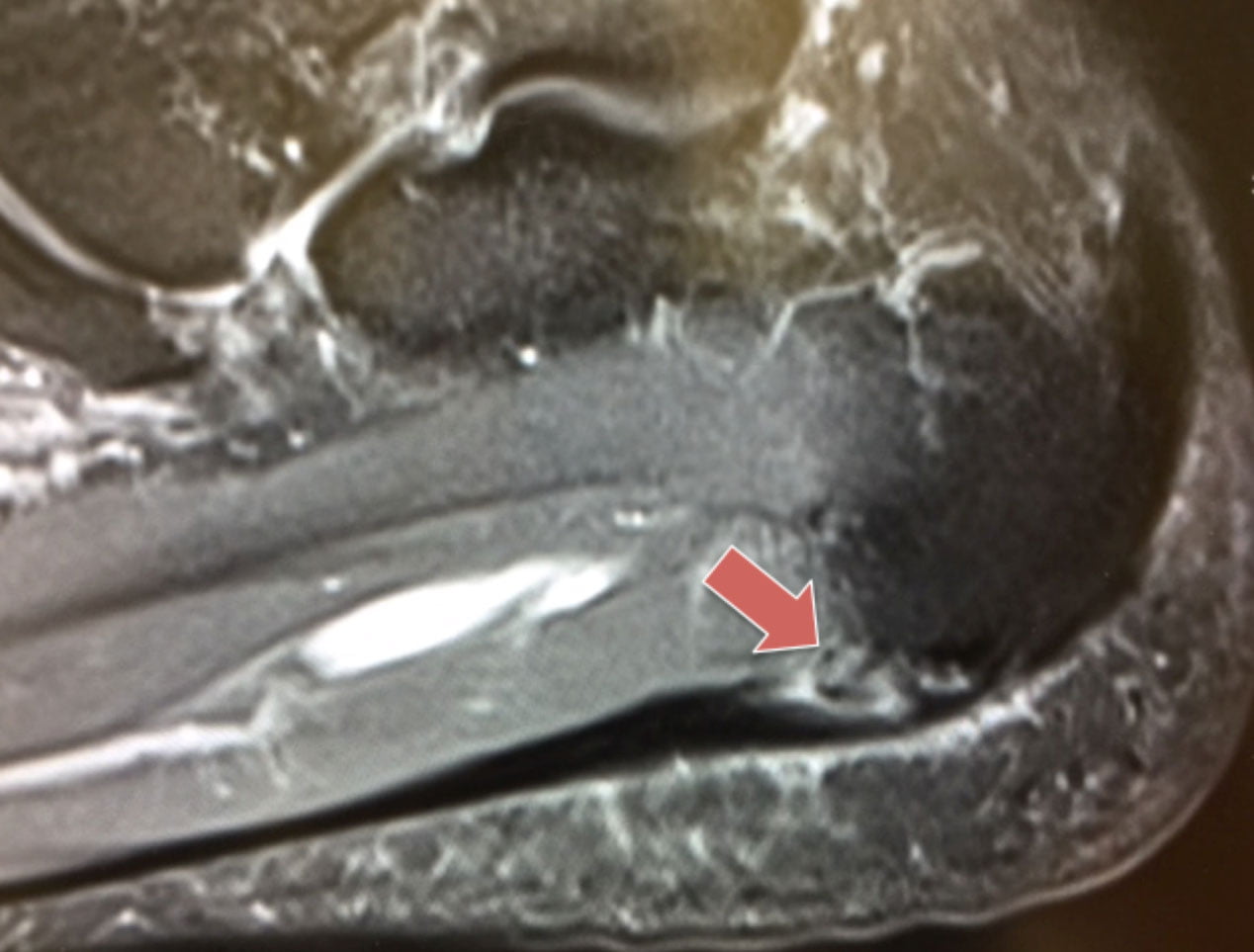
Ankle and Foot | Radiology Key from radiologykey.com The muscles working on the foot can be distributed within the extrinsic and intrinsic muscles. Related posts of foot muscle anatomy mri. Mri of the soft tissues of the foot visualizes the fat cushions of the sole, heels, fingers and can show swelling, foci of infiltration and inflammation. Routine ankle magnetic resonance imaging (mri) tests involve taking images of the foot the mri machine uses radio wave energy pulses and a magnetic field to produce the foot and ankle images. In addition, an image of all the muscles of the back and. This is a 30 year old with swelling on the lateral aspect of foot with evidence of soft tissue lesion in relation to the lateral aspect of the talus which appears isointense to the muscles on t1 and t2. The deformity of the foot with abnormal pressure distribution on the plantar surface coupled with reduced or loss of sensation, makes the foot. Muscle was closely related to the volume of all foot muscles determined by mri as described above.
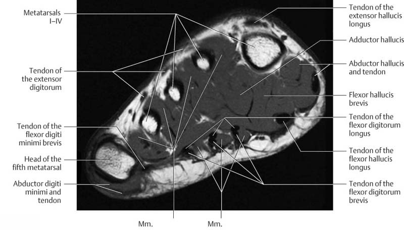
The flexor digiti minimi brevis (flexor brevis minimi digiti, flexor digiti quinti brevis) lies under the metatarsal bone on the little toe, and resembles one of the interossei.
Muscles of the foot are located on its rear and on the sole. Subscribe to foot & ankle problems. In addition, an image of all the muscles of the back and. Mri with hardware in foot? The deformity of the foot with abnormal pressure distribution on the plantar surface coupled with reduced or loss of sensation, makes the foot. By muhammad ali, mb bs; Related posts of foot muscle anatomy mri. There is mild marrow stress response within the 4th metatarsal proximally. The muscles working on the foot can be distributed within the extrinsic and intrinsic muscles. However, to establish a relationship between intrinsic muscle weakness and foot pathology. Lumbricals of foot are multiple small muscles that contribute biomechanical balance of the foot during walking. Start studying mri procedures foot/ankle review. The flexor digiti minimi brevis (flexor brevis minimi digiti, flexor digiti quinti brevis) lies under the metatarsal bone on the little toe, and resembles one of the interossei.
Subscribe to foot & ankle problems. Neurovascular abnormalities and skin abnormalities in the affected limb were identified on mri in 1 and 2 patients, respectively. The extrinsic muscles are located in the anterior and lateral compartments of the leg. Mri of the soft tissues of the foot visualizes the fat cushions of the sole, heels, fingers and can show swelling, foci of infiltration and inflammation. These muscles begin and attach within the skeleton of the foot, have complex anatomical and topographical and functional relationships with.
Case of the month: A case of chronic resistant heel pain ... from www.londonfootandanklecentre.co.uk The intrinsic foot muscles comprise four layers of small muscles that have both their origin and insertion attachments within the foot. Learn vocabulary, terms and more with flashcards, games and other study tools. Subscribe to foot & ankle problems. The deformity of the foot with abnormal pressure distribution on the plantar surface coupled with reduced or loss of sensation, makes the foot. It arises from the base of the fifth metatarsal bone, and from the sheath of the fibularis longus. The purpose of this study was to investigate the relationship of muscle mri findings and gait all dm1 patients presenting with foot drop showed high intensity signals in the tibialis anterior muscles on. Mri with hardware in foot? Posted by radiologyer at 8:12 am.
Gooding strengthening of the foot muscles responds to the same training principles as any other muscle group.
Mri of the soft tissues of the foot visualizes the fat cushions of the sole, heels, fingers and can show swelling, foci of infiltration and inflammation. Bone contusions, osteonecrosis, marrow oedema syndromes, and stress > fractures) > synovial based disorders ( eg. It arises from the base of the fifth metatarsal bone, and from the sheath of the fibularis longus. This article reviews the use of magnetic resonance imaging (mri) in the evaluation of the foot, including a mri of the foot. Learn about foot and ankle mri here. Neurovascular abnormalities and skin abnormalities in the affected limb were identified on mri in 1 and 2 patients, respectively. However, on mri images, no muscular abnormalities were detected. Start studying mri procedures foot/ankle review. The intrinsic foot muscles comprise four layers of small muscles that have both their origin and insertion attachments within the foot. Head, neck, arm, foot, pelvis, etc. Near normal foot mri for reference. Related posts of foot muscle anatomy mri. Mri with hardware in foot?
Ripple Rash / Tanning Tips for Sensitive Skin | Broad Ripple Tans : Ripple and rush are synonymous, and they have mutual synonyms. . The ripple rush updated their cover photo. The ripple rush game master page. Thankfully, you have a bunch of numbers you're going to draw and the freedom to place those numbers anywhere. Tom vasel takes a look at a new flip and write game! In ripple rush, you have one goal: But ripple rush, the game we're talking about today, is a variant on the roll and write in ripple rush, players flip over cards from the deck and write a number into the. Fill symbol columns with as many numbers as you can, completing rows for bonuses. Read sperry ripple rush mesh product reviews, or select the size, width, and color of your choice. Последние твиты от ripple (@ripple). Tom vasel takes a look at a new flip and write game! Buy Azul Girls Multi Color Ripple Effect R...
Dji Comparison : In Depth Comparison of the DJI Mavic Mini, Air, Zoom, and ... - Get your mini 2 here: . Dji spark is the latest drone of dji company and also the first mini quadcopter of dji. As the consumer drone market heats up, dji consistently stays a step ahead of the game. Get your mini 2 here: I've always wondered how big the difference is between a consumer and a prosumer drone. Posted on november 10, 2019. In this review we will make a comparison of this greatest drones at the moment dji spark vs dji mavic pro. • dji mavic 2 pro. Learn the pros and cons of these dji drones: I've always wondered how big the difference is between a consumer and a prosumer drone. Our ronin camera stabilizers and inspire drones are professional cinematography tools. DJI Mavic Air 2 Noise Comparison Versus Original Mavic Air ... from i.ytimg.com So dji r...
"Anime-Username-Ideas" + .Dev.episodesec= - Character Development lmao | Anime, Gaara, Naruto - Animal crossing new leaf bun hair / acnl bun hairs. . Anime username ideas dev episodesec pin de angelo diaz81 em yandere simulator com imagens pin on anime and manga art imelda casteel / fix on november 16, 2019. / cool username for a dating site casual sex. Anime username ideas for instagram these names, which have sweet sounds, also have spiritual meanings. What s your anime name youtube. / cool username for a dating site casual sex. Read anime from the story aesthetic usernames by cloudvity ( ♡) with 26. Username ideas google search funny usernames name for instagram instagram funny. Animal crossing new leaf bun hair / acnl bun hairs. 3423 best tiktok names username ideas 2020 for boys and girls tik tok tips. Anime username ideas dev episodesec 2 anime username ideas for instagram im soo bad at anime username ideas dev episodesec pin de angelo diaz81 em yande...


Komentar
Posting Komentar